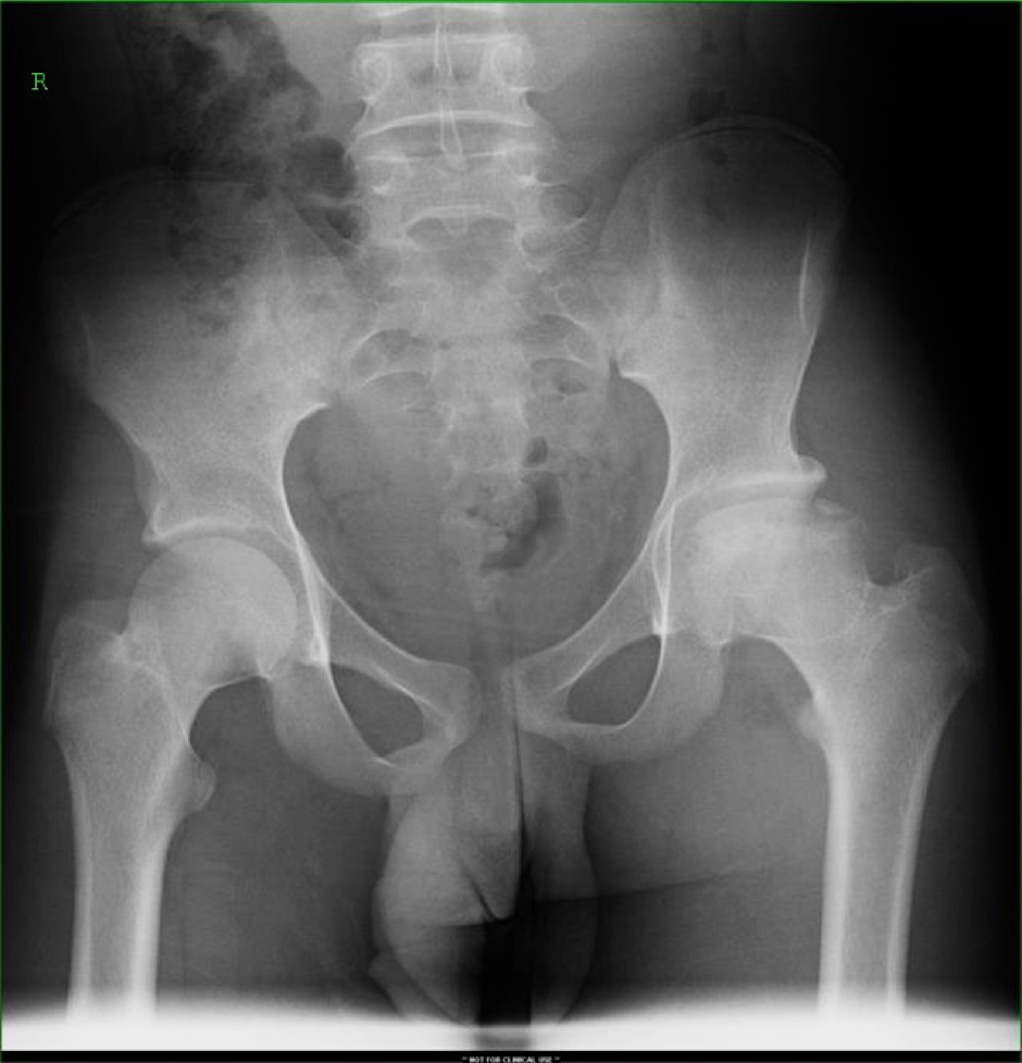
Jamaican male with hip pain
This is my own work and is provided here as an example of how I perform an in-depth image critique but this is not a ‘this is how it must be done’ example.

Clinical history
Jamaican male with hip pain
Request justification
Lack of availability of clinical information makes it difficult to accurately correlate radiographic findings with a definitive diagnosis. It is the responsibility of the referring clinicians to provide accurate information (Van Borsel et al., 2015). In the radiology report, the referring clinicians expect a diagnosis to explain the patient’s symptoms but in exchange, the clinicians have a duty to provide the radiologist with an unequivocal clinical question – which is not seen in this example (Van Borsel et al., 2015). From my experience, the following information should have been included with this request:
– the patient’s age (to cull the common
differentials)
– any recent trauma to the pelvic area
– lifestyle choices that might explain the hip pain (e.g. active in sport
activities)
– previous medical history (e.g. previously diagnosed DDH or AVN)
– current medical treatments (e.g. chemotherapy, radiation therapy)
However, the rationale for the pelvis x-ray given the clinical history is justified. According to Murphy (2017) pelvic radiographs are performed for generalised hip pain. But, a definitive diagnosis may not be possible.
Image quality
A pelvic x-ray is presented with patient identification removed for confidentiality and privacy. A digital right side x-ray marker is seen in the upper corner of the patient’s right side. A true collimation border is seen inferiorly and three post-processed borders are seen framing the image. Using these borders as a guide, the central ray (CR) was directed at roughly the midpoint of the anterior-superior-iliac-spine and pubis-symphsis line. This is the level that the CR should be centred (Bontrager et al., 2014; Knipe et al., 2017).
The distal aspect of L3 is seen superiorly, the superior third of the proximal femori inferiorly and skin edges laterally. There is no unnecessary anatomy included in this radiograph (Bontrager & Lampignano, 2014; Knipe & Murphy, 2017).
In terms of radiographic density, this image has been marginally over-exposed because skin edges of the iliac bones are not seen, suggesting that a high mAs was selected. This is a high contrast radiograph with visualisation of the cortical outlines and trabeculae suggesting that appropriate kVp was selected to demonstrate the pelvis anatomy.
The visualised obturator foramens are equal in size suggesting that the patient’s mid-sagittal plane was parallel with the table top and the bilateral anterior superior iliac spines were also equidistant from the table top. Cortical outlines are sharp and clear – therefore, no patient motion. No removable artefacts are identified.
Radiological description of pathology
Left hip
Femoral head has an irregular contour, demonstrates diffuse patchy sclerosis and subchondral lucency. The femoral head shows sign of collapse and fragmentation. Tracing Letournel’s lines, the acetabular roof is flattened, the anterior and posterior acetabular walls are preserved, and the posterior and anterior columns are intact. No other fractures or avulsions identified.
Right hip
The right hip is radiographically normal in appearance with normal contours. No fractures or avulsions identified.
Both hips
Both femoral heads are enlocated with adequate acetabular coverage (lateral center-edge angle >25 degrees) (Nemattalla, 2015). The acetabular fossa is projected medial to the ilioischial line bilaterally (left > right) – indicating coxa profunda (Kang et al., 2017).
Using the inter-femoral head centre distance (femoral head centre to centre) reference of 15.8 cm for males (Mullaji et al., 2010), the sacroiliac joints (2 mm, bilaterally) and symphysis pubis joint space (5 mm) are extrapolated to be within normal limits (Weerakkody et al., 2018).
Diagnosis
Given the imaging findings, differentials include:
- Avascular necrosis (osteonecrosis) of the femoral head
- A femoral neck fracture
- Legg-Calve-Perthes disease
The patient’s age can be estimated by the presence of incomplete fusion of the iliac ossification centre and Risser’s sign. The radiographic signs suggest that the patient is at Risser Stage 4 putting the patient at 16-17 years of age (Mayet et al., 2010).
The patient’s relatively older presentation (16-17 years) suggests that the pathology demonstrated is unlikely Legg-Calve-Perthes disease which typically has peak presentation at 5-6 years (Niknejad et al., 2018). The lack of trauma implies that traumatic femoral neck fractures can be excluded but pathological fractures cannot be entirely excluded.
The patient is of Jamaican decent and by association African ancestry, sickle cell disease immediately comes to mind (Martí-Carvajal et al., 2016). Radiopaedia.com article on skeletal manifestations of sickle cell disease puts osteonecrosis as a radiographic feature of sickle cell disease (Bell et al., 2018).
In the absence of clinical guidance, radiographic features suggest the diagnosis of osteonecrosis of the femoral head. This may be idiopathic in nature or secondary to an undiagnosed cause like sickle cell disease. Coxa profunda is also appreciated (left > right).
Further imaging
According to Knipe (2018) MRI is the most sensitive modality and in this case study, should be the next imaging choice for further evaluation of the left hip. Bone scintigraphy should also be utilised if the patient has known risk factors like sickle cell disease to assess for multiple sites of involvement.
References
Bell, D. J., & Stanislavsky, A. (2018). Sickle cell disease (skeletal manifestations). Retrieved from https://radiopaedia.org/articles/sickle-cell-disease-skeletal-manifestations-1
Bontrager, K., & Lampignano, J. (2014). Bontrager’s handbook of radiographic positioning and techniques
Kang, O., & Knipe, H. (2017). Coxa profunda. Retrieved from https://radiopaedia.org/articles/coxa-profunda
Knipe, H. (2018). Avascular necrosis. Retrieved from https://radiopaedia.org/articles/avascular-necrosis
Knipe, H., & Murphy, A. (2017). Pelvis (AP view). Retrieved from https://radiopaedia.org/articles/pelvis-ap-view-1
Martí-Carvajal, A. J., Solà, I., & Agreda-Pérez, L. H. (2016). Treatment for avascular necrosis of bone in people with sickle cell disease. Cochrane Database of Systematic Reviews(8). doi:10.1002/14651858.CD004344.pub6
Mayet, Z., Lukhele, M., & Mohammed, N. (2010). Risser sign – Trends in South African population. SA Orthopaedia Journal.
Mullaji, A., Shetty, G. M., Kanna, R., & Sharma, A. (2010). Variability in the range of inter-anterior superior iliac spine distance and its correlation with femoral head centre. A prospective computed tomography study of 200 adults. Skeletal Radiology, 39(4), 363-368. doi:10.1007/s00256-009-0791-x
Murphy, A. (2017). Pelvis series. Retrieved from https://radiopaedia.org/articles/pelvis-series
Nemattalla, W. (2015). Normal acetabular rims and Wiberg angle. Retrieved from https://radiopaedia.org/cases/normal-acetabular-rims-and-wiberg-angle
Niknejad, M. T., & Gaillard, F. (2018). Perthes disease. Retrieved from https://radiopaedia.org/articles/perthes-disease
Van Borsel, M. D., Devolder, P. J. D., & Bosmans, J. M. L. (2015). Software solutions alone cannot guarantee useful radiology requests. Acta Radiologica, 57(11), 1366-1371. doi:10.1177/0284185115588225
Weerakkody, Y., & Jones, J. (2018). Pelvic Radiograph (an approach). Retrieved from https://radiopaedia.org/articles/pelvic-radiograph-an-approach
0 comments on “Image critique example – pelvis” Add yours →